
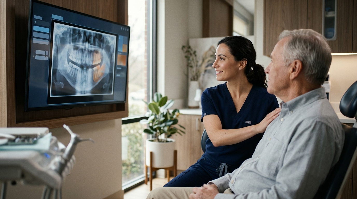
Digital X-rays detect bone problems in oral cancer by revealing subtle changes in jaw density and structure that are invisible to the naked eye. At Tooth Doc Family Dentistry in Herndon, VA, we use advanced imaging to identify these early signs during routine screenings. This technology allows us to spot abnormalities before they cause pain or significant damage, ensuring timely and effective treatment for our patients.
How Digital Imaging Reveals Bone Changes
Digital X-ray technology is a method of capturing high-resolution images of the jaw and teeth using low-dose radiation. Unlike traditional film, digital sensors convert radiation into electronic data that can be magnified and analyzed on a screen. This clarity is critical for detecting bone loss or irregularities associated with oral cancer. The American Dental Association emphasizes that early detection significantly improves survival rates, making precise imaging tools essential in modern dentistry.
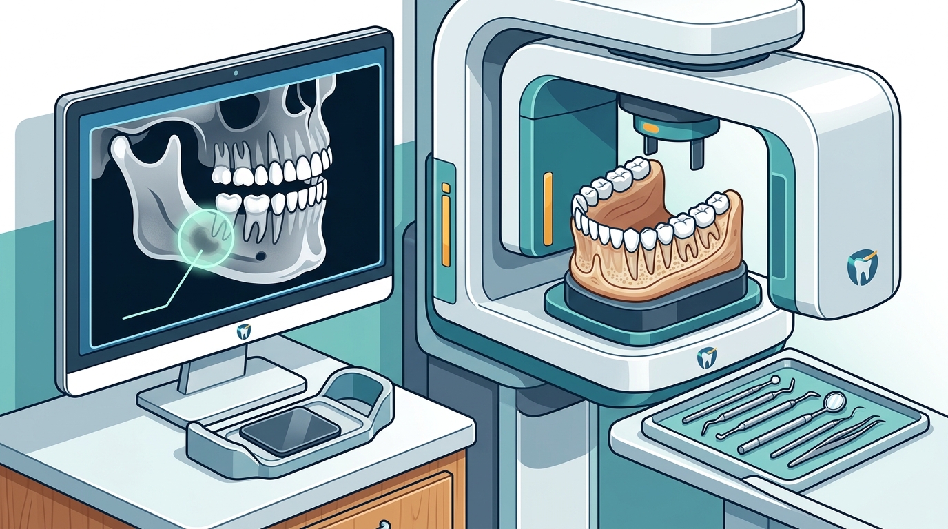
When cancer affects the jawbone, it often causes a process called osteolysis, where the bone tissue breaks down. Digital X-rays can show these areas of decreased density as darker patches or irregular shapes. By comparing current images to previous records, our team can track changes over time. This longitudinal approach helps distinguish between normal aging or wear and pathological bone destruction.
Identifying Specific Bone Abnormalities
Oral cancer does not always present as a visible sore on the gums or tongue. Sometimes, the disease begins in the bone itself or spreads to the jaw from nearby tissues. Digital X-rays help identify specific signs such as moth-eaten bone patterns, which indicate aggressive bone loss. We also look for periosteal reactions, where the outer layer of the bone thickens in response to underlying disease.
One concrete example of a detectable abnormality is a radiolucent lesion, which appears as a dark area on the X-ray. While many radiolucent lesions are benign, such as cysts, their presence warrants further investigation. If a lesion has irregular borders or causes expansion of the jawbone, it raises the suspicion of malignancy. Our team uses these visual cues to determine when a biopsy or referral to a specialist is necessary.
Advantages of Digital Over Traditional Film
Traditional film X-rays have limitations in contrast and resolution that can obscure subtle bone changes. Digital imaging offers superior dynamic range, allowing us to adjust brightness and contrast to highlight specific areas of interest. This flexibility is particularly useful when examining the mandible or maxilla for early signs of cancer. The ability to zoom in on a specific region without losing image quality is a significant advantage in diagnostic accuracy.
Furthermore, digital X-rays can be integrated with software that enhances specific tissue densities. This technology helps differentiate between soft tissue masses and bone involvement. For patients at higher risk, such as those with a history of tobacco use or heavy alcohol consumption, this level of detail is invaluable. It ensures that no subtle bone change goes unnoticed during a routine checkup.
The Role of Screening at Tooth Doc
At Tooth Doc Family Dentistry, we integrate digital X-rays into every comprehensive oral cancer screening. We understand that many patients are unaware of the link between bone health and oral cancer. By explaining what we see on the X-ray, we empower patients to make informed decisions about their care. Our conservative approach means we only recommend further testing when the imaging results suggest a genuine concern.
We also use digital records to monitor patients who have had previous oral health issues. If a patient has a history of jaw trauma or prior cancer, we pay extra attention to those areas during imaging. This personalized monitoring helps us catch any recurrence or new development early. Our goal is to provide peace of mind through thorough and transparent diagnostic practices.
Next Steps After Detection
If a digital X-ray reveals a suspicious bone abnormality, the next step is usually a clinical examination and potentially a biopsy. We do not diagnose cancer based on an X-ray alone, as imaging provides clues rather than a definitive answer. However, the X-ray guides the dentist on where to look and what to test. This targeted approach reduces the need for unnecessary invasive procedures.
For patients referred to an oral surgeon or oncologist, our digital records provide a clear baseline for treatment planning. The surgeon can use the X-ray images to plan the extent of any necessary surgery. This continuity of care ensures that the patient receives the most appropriate treatment from the moment of detection. We support our patients through every step of this process, from initial screening to follow-up care.
Key Takeaways
- Digital X-rays detect bone problems by revealing changes in jaw density and structure.
- Early detection of bone abnormalities can significantly improve oral cancer survival rates.
- Specific signs like moth-eaten patterns and radiolucent lesions indicate potential malignancy.
- Regular screenings at Tooth Doc Family Dentistry include comprehensive digital X-ray analysis.
- Imaging guides clinical decisions, ensuring that biopsies and referrals are targeted and necessary.
Frequently Asked Questions
Can digital X-rays detect oral cancer in the bone?
Yes, digital X-rays can detect changes in the jawbone that may indicate oral cancer. These changes often appear as areas of decreased density or irregular bone structure. While an X-ray alone cannot confirm cancer, it is a critical tool for identifying when further testing is needed.
How often should I get a digital X-ray for cancer screening?
Most patients receive digital X-rays during their routine dental exams, typically every six months to a year. The frequency may vary based on your individual risk factors and dental history. Your dentist will recommend the appropriate schedule for your specific needs.
Are digital X-rays safe for frequent use?
Digital X-rays use significantly less radiation than traditional film X-rays. The dose is low enough that the benefits of early detection far outweigh the minimal risk of radiation exposure. We follow strict safety protocols to ensure patient protection during every imaging session.
What happens if my X-ray shows a bone abnormality?
If an abnormality is detected, we will perform a thorough clinical examination to correlate the imaging findings with physical symptoms. We may recommend a biopsy or refer you to a specialist for further evaluation. This step ensures an accurate diagnosis and appropriate treatment plan.
Does Tooth Doc Family Dentistry use digital X-rays?
Yes, we use state-of-the-art digital X-ray technology for all our patients. This allows us to provide high-quality images that help detect bone problems and other oral health issues early. You can view your digital records on our office screens during your visit.
Conclusion
Digital X-rays are a vital component of early oral cancer detection, particularly for identifying bone abnormalities that are invisible to the eye. By leveraging this technology, we can provide our patients in Herndon with the best chance of early diagnosis and successful treatment. If you have not had a recent dental exam, we encourage you to schedule an appointment with us. To plan your visit, today.